
Spiny Dogfish Dissection: A Step by Step Guide
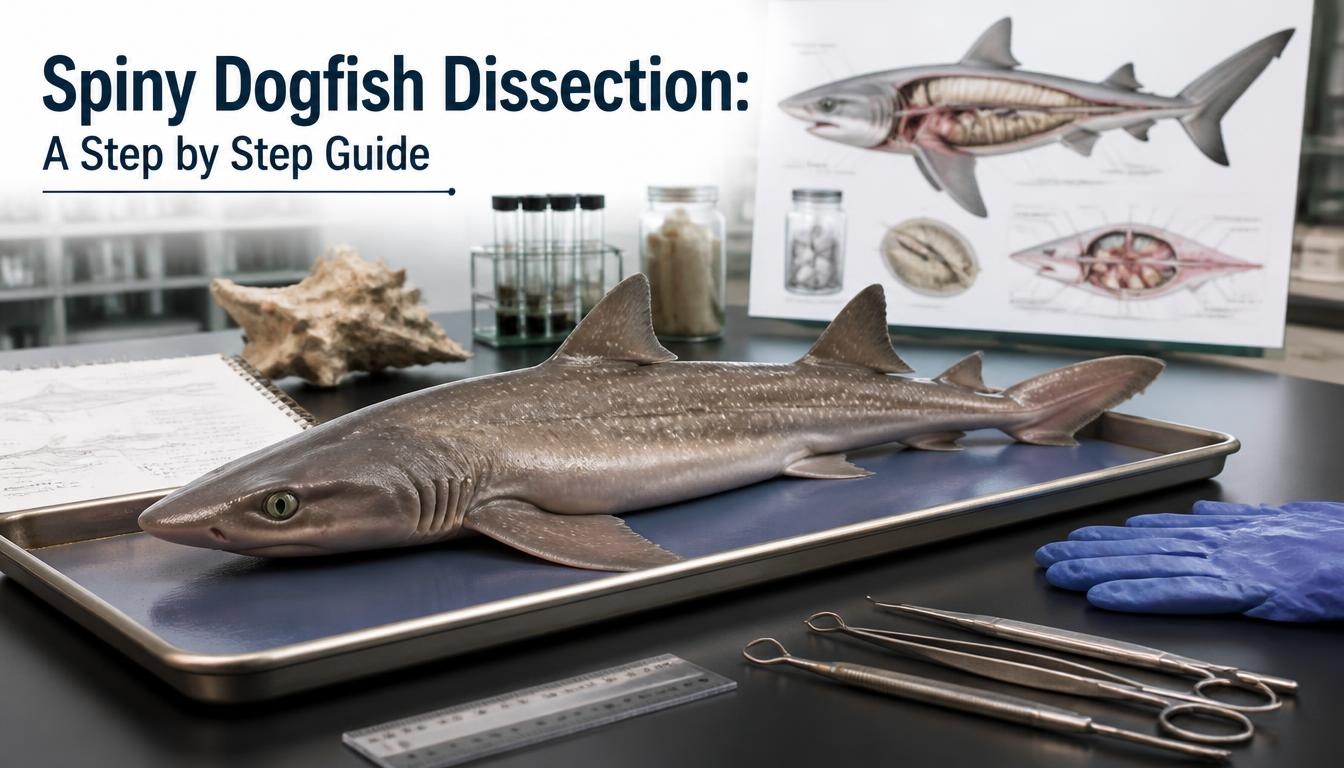
A spiny dogfish dissection is one of the most common introductory shark anatomy labs taught in biology classrooms, and for good reason: the spiny dogfish (Squalus acanthias) is small, widely available, and displays nearly every major structural feature found across cartilaginous fish. This guide walks through the full dissection process step by step, from external identification to the internal organ systems you'll actually be asked to locate and label.
What You'll Need Before You Start
A standard spiny dogfish dissection requires a preserved specimen, a dissection tray, dissecting scissors, a scalpel, forceps, dissecting pins, and protective gloves. Work in a well ventilated space, since preservative fumes (typically formalin based) can be irritating with prolonged exposure, and keep your specimen moist with preservative solution or water between dissection sessions to prevent it from drying out and stiffening.
Step 1: External Anatomy Identification
Before making any incision, spend time identifying external features, since several are diagnostic for the species and provide useful orientation for the dissection to come. Spiny dogfish get their name from a sharp spine positioned just in front of each of their two dorsal fins a genuine defensive feature, so handle the dorsal area carefully even on a preserved specimen. Count the gill slits along the side of the head; spiny dogfish have five, a standard number across most shark species. Just behind and slightly above each eye, look for a small opening called the spiracle, a modified gill slit used for drawing water over the gills while the shark is resting or feeding on the bottom. Finally, check the pelvic fins: males have a pair of modified claspers extending from the pelvic fins, used in reproduction, while females lack this structure entirely the fastest way to determine your specimen's sex before you begin.
Step 2: Making the Initial Incision
Place the specimen ventral (belly) side up in the dissection tray, pinning the fins outward if needed to keep it stable. Using scissors, make a shallow incision starting just anterior to the cloaca (the posterior opening used for waste and reproduction) and extend it forward along the midline toward the pectoral girdle, cutting through skin and muscle without puncturing the organs beneath shallow, controlled cuts matter far more here than speed. From that midline cut, make two lateral incisions angling outward toward each pectoral fin, creating a set of flaps that can be pinned back to expose the full body cavity, similar to opening a book.
Step 3: Exposing the Body Cavity
Once the flaps are pinned back, you should see the body cavity (coelom) and the major organs held loosely within it, surrounded by a thin lining called the peritoneum. Take a moment to note the overall layout before removing or repositioning anything the liver's large, paired lobes are typically the most immediately visible structure, occupying much of the anterior cavity.
Step 4: The Digestive System
Trace the digestive tract from the esophagus, a short muscular tube leading from the mouth, down into the J shaped stomach. Beyond the stomach, the small intestine leads into the most distinctive digestive structure in a shark's body: the spiral valve intestine, a short but internally elaborate section of gut lined with a corkscrew shaped fold that dramatically increases surface area for nutrient absorption within a compact length a structural solution cartilaginous fish evolved instead of the long, coiled intestines found in most bony fish. Locate the liver's large lobes (which also serve a buoyancy function in sharks, since they lack a swim bladder), the small pancreas nestled near the first loop of intestine, and the gallbladder, a small greenish sac connected to the liver that stores bile.
Step 5: The Circulatory System
The shark heart sits anterior to the liver, near the gill region, and is structured differently from a bony fish heart in one key way worth noting: it includes four chambers in sequence the sinus venosus, atrium, ventricle, and conus arteriosus arranged to move blood in a single direction toward the gills. Identifying each chamber in sequence, rather than as a single compact structure, is one of the most commonly tested elements of a spiny dogfish dissection.
Step 6: The Reproductive & Urogenital System
Reproductive structures differ significantly by sex, which is exactly why identifying sex early in the dissection (Step 1) matters. In females, paired ovaries sit near the anterior body cavity, connected to oviducts leading toward the cloaca; spiny dogfish are ovoviviparous, meaning eggs develop and hatch internally before live birth, with one of the longest gestation periods documented in any vertebrate, commonly cited between roughly 18 and 24 months. In males, paired testes connect to the claspers observed externally in Step 1, which are used to transfer sperm during mating. Both sexes have paired kidneys running along the dorsal body wall, connected to the urogenital system.
FAQ
What is the most distinctive internal structure in a spiny dogfish dissection?
The spiral valve intestine a compact section of intestine lined with a corkscrew shaped fold that increases nutrient absorption surface area, replacing the long coiled intestine found in most bony fish.
How can you tell if a spiny dogfish specimen is male or female before dissecting?
Check the pelvic fins. Males have a pair of claspers, rod like structures used in reproduction; females lack this structure entirely.
How many gill slits does a spiny dogfish have?
Five, a standard number found across most shark species, visible externally along the side of the head.
What is the spiracle, and where is it located?
A modified gill slit positioned just behind and slightly above each eye, used to draw water over the gills, particularly useful when the shark is resting or feeding near the bottom.
Why does a shark's liver matter so much in dissection?
Beyond its digestive role, the liver serves a buoyancy function in sharks, since they lack a swim bladder its large size in the body cavity reflects this dual purpose.
How long is the gestation period for spiny dogfish?
Commonly cited between 18 and 24 months, one of the longest gestation periods documented in any vertebrate, tied to their ovoviviparous reproductive strategy.
What safety precautions matter most during a spiny dogfish dissection?
Handle the area near the dorsal fins carefully, since each carries a genuine spine even on a preserved specimen, and work in a well ventilated space due to preservative fumes.
Conclusion
A spiny dogfish dissection walks through nearly every major structural adaptation that defines cartilaginous fish the spiral valve intestine solving digestion in a compact body, a four chambered heart arranged for one directional blood flow, and a reproductive strategy built around one of the longest gestation periods of any vertebrate. Working through each system methodically, in the order outlined here, makes it far easier to locate and understand every structure you'll be asked to identify. For more on shark and fish anatomy generally, see our guide on what fisheries science actually studies.